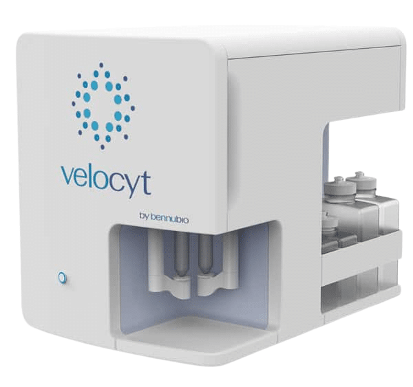
The Velocyt® delivers breakthrough technological innovation in flow cytometry for cancer research and drug development. It’s the world’s first ever rapid, large particle analysis system and is poised to transform how fundamental life science research is conducted.
Parallel Streams
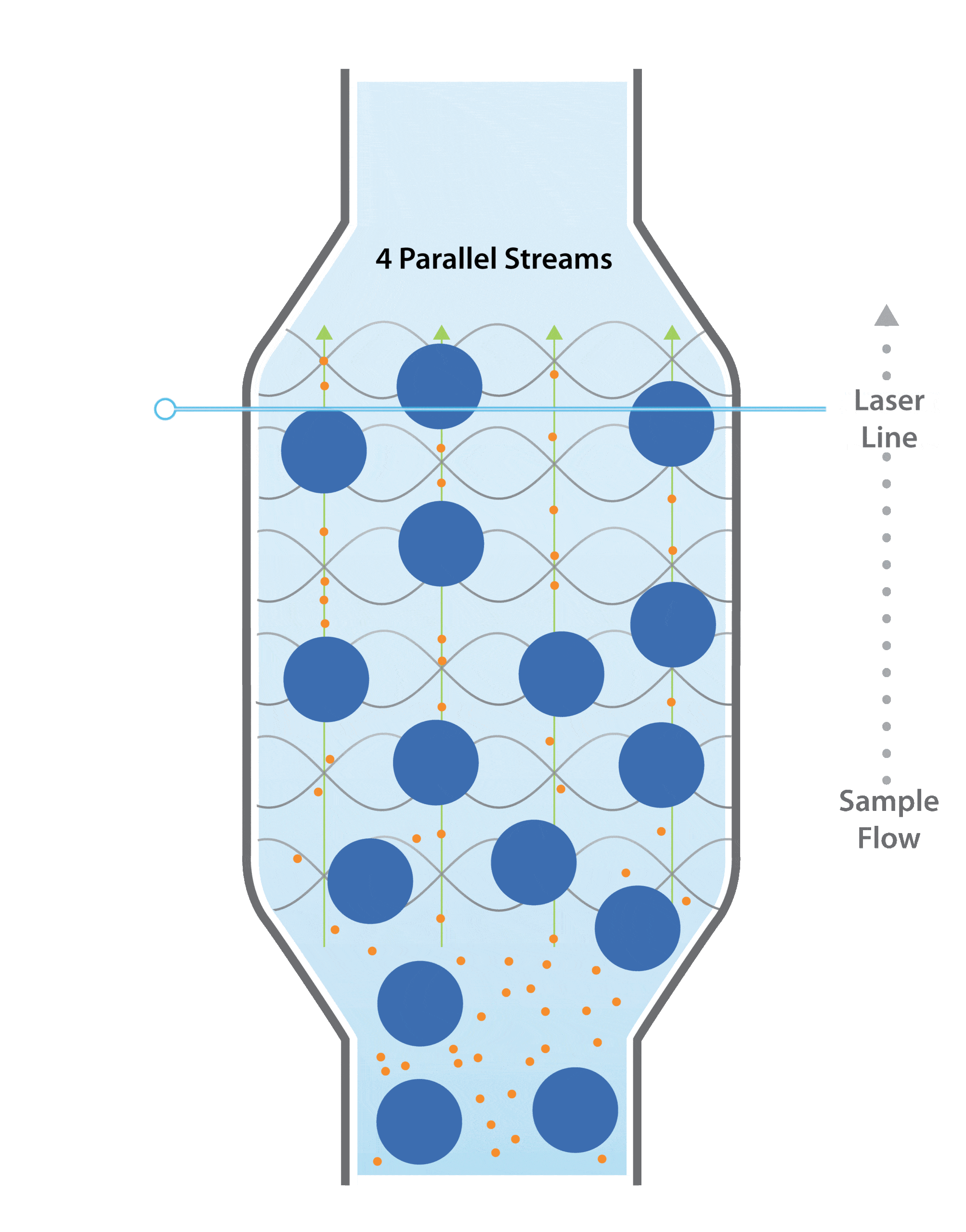
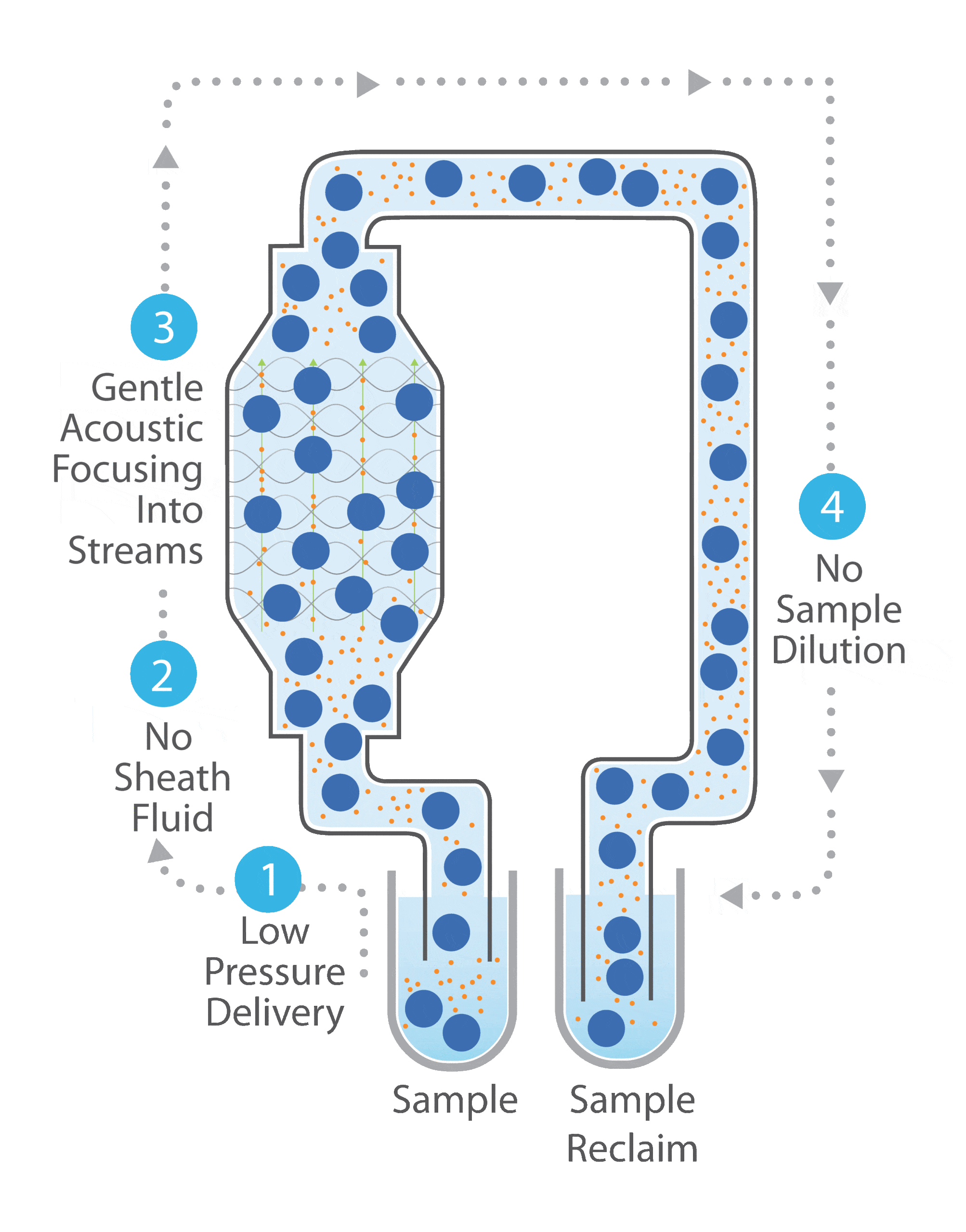
The Velocyt® uses a patented system to generate parallel flow streams with acoustic standing waves and simple laminar flow through the flow cell
- Acoustic standing waves are driven at megahertz frequencies in a flow channel that has acoustically reflective walls.
- This acoustic force drives particles that have higher density and that are less compressible than the surrounding fluid to the “pressure nodes”.
- The frequency of the standing waves can be set such that a known number of streams is generated across the channel.
- Vertical positioning is achieved using wide shallow channels that enable the lift forces generated by flowing fluid to move particles into a vertically centered position.
- Particles flow through one or more optical sensing regions by simply pumping the complete sample through the flow cell.

Large Particle Analysis
Our large flow channel width and depth allows The Velocyt® to accommodate particles as large as 300 µm in diameter. Combined with our multistream detection approach, this enables large particle analysis more efficiently and at high spatial resolution.
Total Sample Recovery
Our acoustic focusing approach does not require sheath fluid, which enables recovery of the unaltered sample. Our high flow rates allow analysis of your entire sample, which is returned to you intact. This is extremely valuable for samples that are costly, otherwise precious or needed for off-line analysis. This also makes The Velocyt® the only flow cytometer capable of simple kinetic analysis of the same, unaltered sample.

Correlated Optical Detection
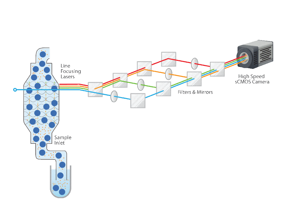
Our patented fluidic approach is coupled with a patented approach to optical interrogation. A line focused laser illuminates across all streams, which constrains the illumination to reduce background while providing sufficient power for resolving dim particles. An extremely fast sCMOS camera images the laser intersection region(s) to record scatter and fluorescence signals from each focused stream simultaneously.

Simple Data Acquisition
Images for each stream are processed into signal pulses and standard flow cytometry parameters are extracted (signal height, width, area, ToF). Data from all streams are integrated into a single data file containing all events, displayed with a custom user interface and available as standard FCS files.
