Watch Seminar Presentation
Watch BennuBio President and CEO, John O'Rourke, give a talk on a simplified approach for rapid analysis of thousands of intact spheroids using The Velocyt® Large Particle Imaging Cytometer as part of a collaboration with Corning.
Ditch The Dissociation: A Simplified Approach for Rapid High Throughput Multiplexed Analysis of Intact 3D Multicellular Spheroids Using The Velocyt® Large Particle Imaging Cytometer
BennuBio White Paper
In this application note, we demonstrate the use of The Velocyt® cytometer for the spheroid workflow using staurosporine treatment as a model drug compound. In this case study, we illustrate Bennubio’s simplified spheroid solution approach (S³) to generate and treat large numbers of uniform spheroids, a simplified sample preparation workflow and the rapid analysis of thousands of intact spheroids, generating flow cytometry, fluorescence imaging and morphological data from each spheroid.
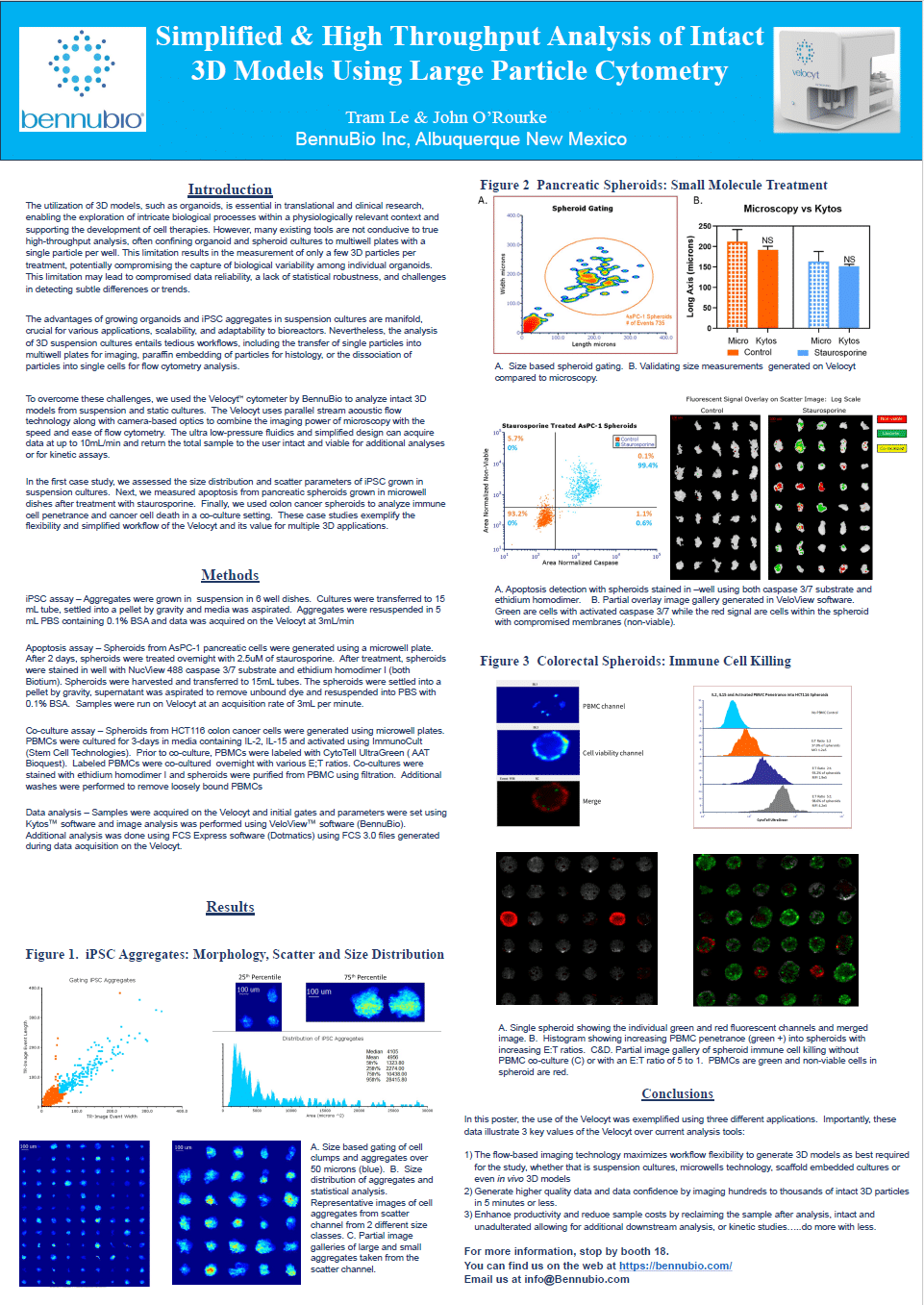
Streamlining 3D Model Analysis: High-Throughput Applications of The Velocyt® Large Particle Imaging Cytometer in Suspension and Static Cultures
In the first case study, we assessed the size distribution and scatter parameters of iPSC grown in suspension cultures. Next, we measured apoptosis from pancreatic spheroids grown in microwell dishes after treatment with staurosporine. Finally, we used colon cancer spheroids to analyze immune cell penetrance and cancer cell death in a co-culture setting.
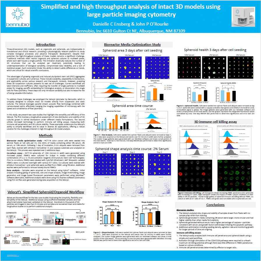
Enhancing 3D Culture Analysis: Media Optimization and Immune Cell Killing Assays Using The Velocyt® Large Particle Imaging Cytometer
In this study, we present two case studies that highlight the versatility and efficiency of The Velocyt®. The first involves a longitudinal assessment of size distribution and viability of 3D cultures grown in stirred bioreactors under different media formulations. The second utilizes microwell technology to perform immune cell killing assays, measuring both immune cell penetration and 3D culture viability.
These examples underscore The Velocyt®’s ability to simplify workflows and its value in diverse 3D applications, offering a robust solution for the challenges inherent in high-throughput 3D model analysis.
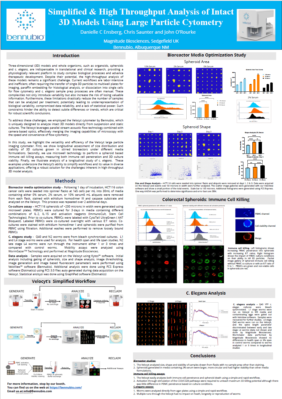
High-Throughput Imaging and Analysis of 3D Biological Models Using The Velocyt® Large Particle Imaging Cytometer
In this study, we present three case studies that highlight the versatility and efficiency of The Velocyt®. The first involves a longitudinal assessment of size distribution and viability of HCT116 3D cultures grown in stirred bioreactors under different serum concentrations. The second utilizes microwell technology to perform immune cell killing assays, evaluating both immune cell penetration and 3D culture viability in co-culture with PBMCs. The third demonstrates The Velocyt®’s application in a longitudinal study of C. elegans, analyzing healthspan, lifespan, and mobility across different life stages.
These examples underscore the The Velocyt®’s ability to simplify workflows and its value in diverse 3D applications, offering a robust solution for the challenges inherent in high-throughput 3D model analysis.
