Overview
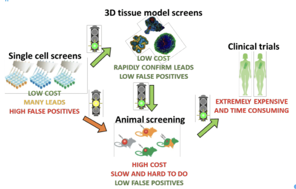
With 90% of drugs failing in clinical trials, research shifted to 3D cell cultures of spheroids and organoids for oncology and disease modeling. 3D cell cultures more closely resemble in vivo cell environments, and provide more precision in drug discovery. And, because they better replicate the in vivo environment, they can reduce the need for animal studies and limit the number of compounds needed to be verified in animal models. The use of 3D cell models in initial and secondary screening steps leads to a lower cost of discovery for many diseases, particularly cancer.
The current methods of 3D cellular analysis are severely limited in terms of particle size and throughput. Specifically, traditional flow cytometers are limited to particles < 50 microns in diameter, while imaging systems and sheath-based large particle flow cytometers analyze far too slowly to be effective. In addition, sheath-based flow cytometers generate significant shear forces that are highly detrimental spheroid health.
The Velocyt analyzes 3D tissue models simply, gently, and rapidly enough to provide the required levels of statistical confidence for the following types of drug discovery studies.
- Drug transport analysis
- Bioavailability studies
- Initial drug targeting screens

